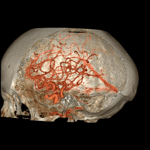
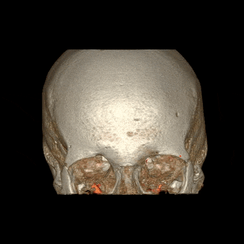
The skull and the Circle of Willis – a structure which supplies blood to the brain and surrounding area.
Computed Tomography (CT) is based on the x-ray principal: as x-rays pass through the body, they are absorbed or attenuated (weakened) at differing levels creating a matrix or profile of x-ray beams of different strength. But while x-rays can only be used to image the outlines of bones and organs, CT creates three-dimensional images of the body one narrow slice at a time.
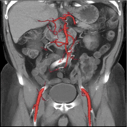
The abdomen and the aorta.
A CT scanner basically looks like a square shaped doughnut fitted with an x-ray tube mounted on one side and the banana shaped detector mounted on the opposite side. As the frame rotates, it takes timely snapshots of whatever or whoever rests inside it. Each time the x-ray tube and detector make a 360° rotation, an image or “slice” has been acquired and by stacking each slice atop another you eventually get a full 3-D image. If all this sounds familiar, it’s because CT is very similar to functioning magnetic resonance imaging (fMRI). Some key differences are that fMRI exploits powerful magnet and pulsing radiowaves, which do not emit potentially harmful radiation, unlike CT.
Where CT shines, however, is in its ability to image tissue inside the body otherwise unapproachable using other methods. All of the GIFs in this post were made from computer images taken using General Electric’s Revolution CT, first introduced in 2013. The device is designed to emit less radiation and provide more comfort. Guts, veins, brains and hearts have now been imaged in the gruesomest detail ever.
Subscribe to our newsletter and receive our new book for FREE
Join 50,000+ subscribers vaccinated against pseudoscience
By subscribing you agree to our
Privacy Policy. Give it a try, you can unsubscribe anytime.
Another view of the Circle of Willis. It is located at the base of the brain and forms a circle of arteries around it.
Another view of the Circle of Willis.
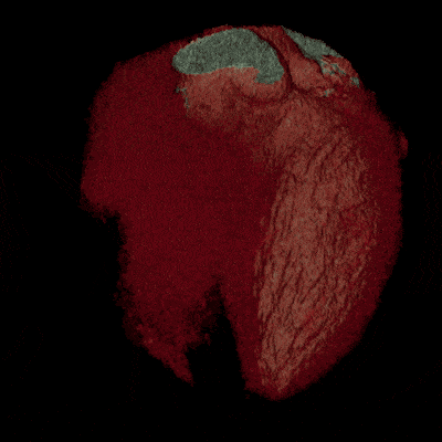
An image of a complete heart in just a single heartbeat.
BONUS: still, high definition images taken with the Revolution CT
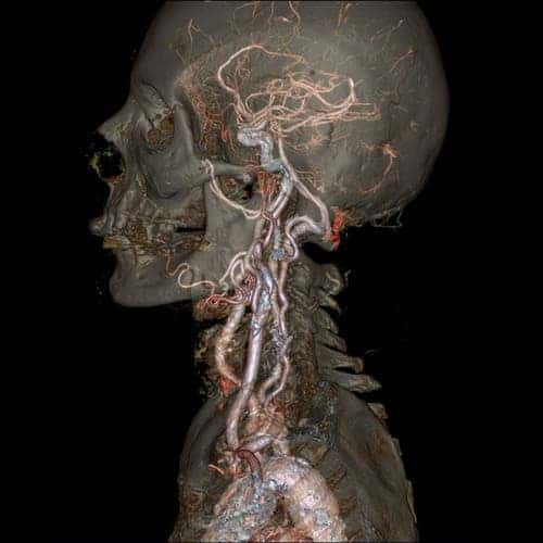
The skull and carotid arteries.
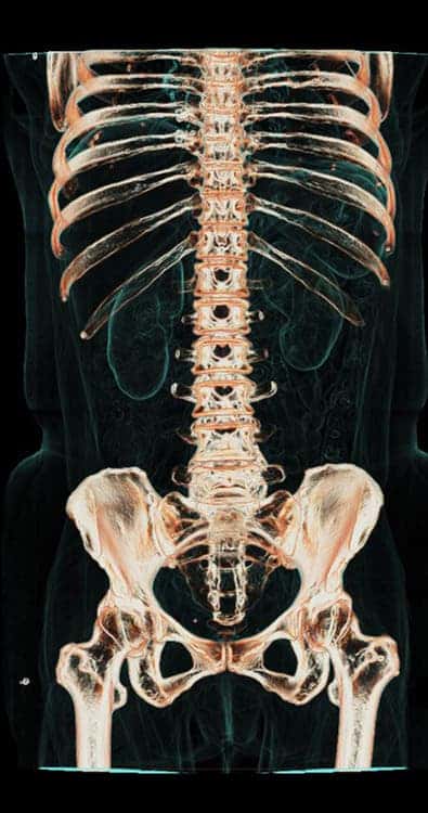
An image of the abdomen and pelvis.
The rib cage, the heart and the chest cavity. The Revolution CT can image the heart in a single heartbeat.
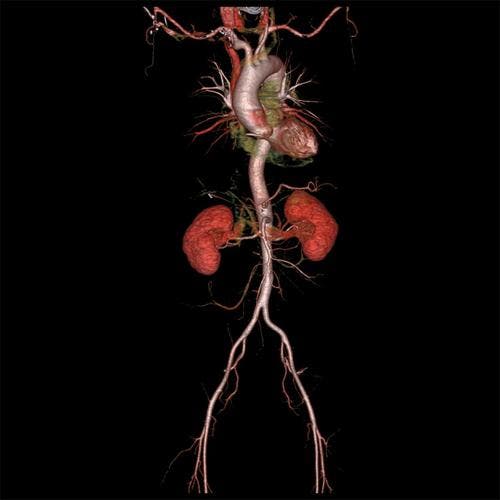
The whole aorta and kidneys.
Original Text (This is the original text for your reference.)
The skull and the Circle of Willis – a structure which supplies blood to the brain and surrounding area.
Computed Tomography (CT) is based on the x-ray principal: as x-rays pass through the body, they are absorbed or attenuated (weakened) at differing levels creating a matrix or profile of x-ray beams of different strength. But while x-rays can only be used to image the outlines of bones and organs, CT creates three-dimensional images of the body one narrow slice at a time.
The abdomen and the aorta.
A CT scanner basically looks like a square shaped doughnut fitted with an x-ray tube mounted on one side and the banana shaped detector mounted on the opposite side. As the frame rotates, it takes timely snapshots of whatever or whoever rests inside it. Each time the x-ray tube and detector make a 360° rotation, an image or “slice” has been acquired and by stacking each slice atop another you eventually get a full 3-D image. If all this sounds familiar, it’s because CT is very similar to functioning magnetic resonance imaging (fMRI). Some key differences are that fMRI exploits powerful magnet and pulsing radiowaves, which do not emit potentially harmful radiation, unlike CT.
Where CT shines, however, is in its ability to image tissue inside the body otherwise unapproachable using other methods. All of the GIFs in this post were made from computer images taken using General Electric’s Revolution CT, first introduced in 2013. The device is designed to emit less radiation and provide more comfort. Guts, veins, brains and hearts have now been imaged in the gruesomest detail ever.
Subscribe to our newsletter and receive our new book for FREE
Join 50,000+ subscribers vaccinated against pseudoscience
By subscribing you agree to our
Privacy Policy. Give it a try, you can unsubscribe anytime.
Another view of the Circle of Willis. It is located at the base of the brain and forms a circle of arteries around it.
Another view of the Circle of Willis.
An image of a complete heart in just a single heartbeat.
BONUS: still, high definition images taken with the Revolution CT
The skull and carotid arteries.
An image of the abdomen and pelvis.
The rib cage, the heart and the chest cavity. The Revolution CT can image the heart in a single heartbeat.
Disclaimer: The translated content is provided by third-party translation service providers, and IKCEST shall not assume any responsibility for the accuracy and legality of the content.


 User Center
 My Training Class
 Feedback


Comments
Something to say?
Log in or Sign up for free